




Shaanxi BLOOM Tech Co., Ltd. is one of the most experienced manufacturers and suppliers of giemsa stain powder cas 51811-82-6 in China. Welcome to wholesale bulk high quality giemsa stain powder cas 51811-82-6 for sale here from our factory. Good service and reasonable price are available.
Giemsa stain powder is a blue purple powder with the chemical formula C14H14ClN3S, CAS 51811-82-6, and a molecular weight of 291.7991. It is easily soluble in equal amounts of methanol and glycerol, forming a blue solution that is flammable. Giemsa pigment was first formulated and applied to biological staining by Gustav Giemsa, a Hamburg chemist and bacteriologist from Germany, in the late 19th century. Its main components include a mixture of Azure II and Azure II Eosin, which enables it to exhibit good selectivity and contrast when staining different biological structures. Applied in other specific studies.

For example, in immunohistochemistry research, Giessen's pigments can be used as auxiliary staining agents to help researchers better observe and analyze the results of antigen antibody reactions; In environmental monitoring, Giemys pigments can be used for staining and counting studies of certain microorganisms; In biochemical experiments, Giemys pigments can also be used for staining and detection of certain enzymes or proteins. As an efficient and multifunctional analytical dye, it has broad application prospects in the fields of biology and medicine. Whether it is genetic research, cytological research, or pathological diacrisis, it is necessary to use Giemys staining technology to reveal the microstructure and morphological characteristics of biological samples.
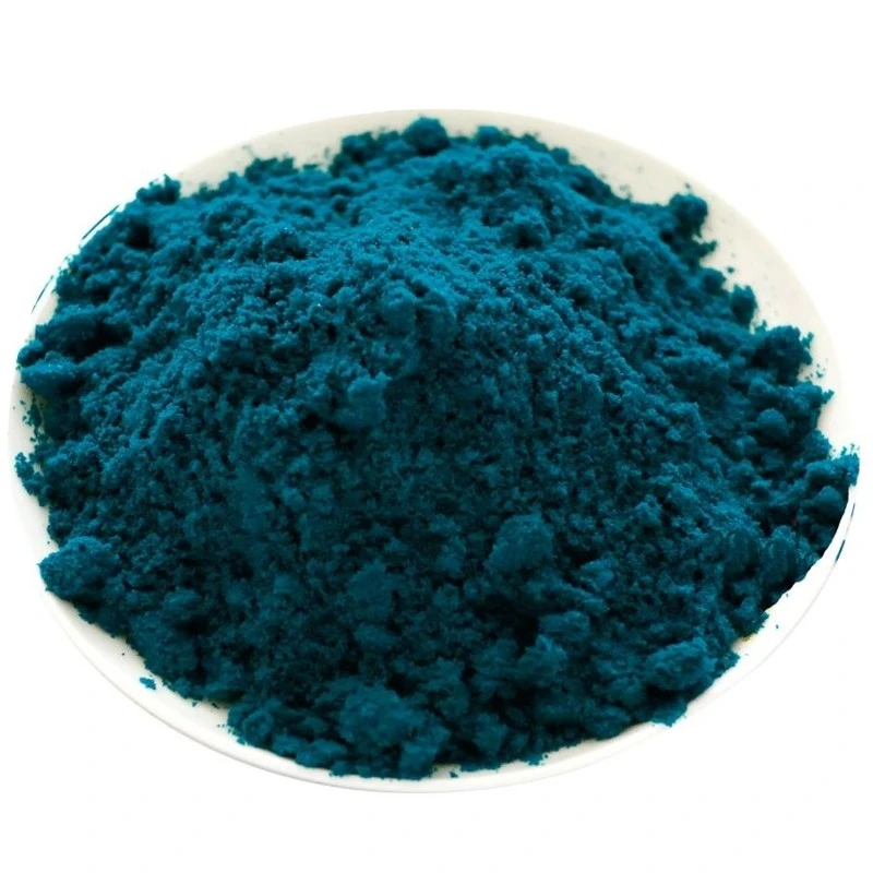

Giemsa stain powder, CAS number 51811-82-6, also known as Giemsa stain, Giemsa dye, or Giemsa colourant solution, is a classic colourant first formulated and applied in the field of biological staining by Gustav Giemsa, a Hamburg chemist and bacteriologist from Germany. Its appearance is a crystalline powder ranging from green to black to dark green, and the solution turns blue when dissolved in methanol and glycerol. The prepared solution is flammable.
The Gi's pigment mainly contains a mixture of Azure II and Azure II eosin. The commercially available dark red Gi's solution consists of Azure B, Azure II eosin, and (alkaline) methylene blue in a mass ratio of 1:12:2, dissolved in glycerol: methanol=5:24 by volume, with a total concentration of 0.6% by mass and a density of 0.89g/cm ³ at 20 ℃. As a neutral colourant, it occupies a pivotal position in biomedical research. Its wide range of applications and powerful functions can be regarded as the "Swiss Army Knife" in the field of biological staining.

1. Original biological staining
The most classic and core use is undoubtedly as an original biological colourant. This application spans across multiple disciplines such as parasitology, microbiology, and hematology, and is an essential foundational tool in laboratories worldwide.
1.1 Blood coating method for detecting malaria parasites
This is the most well-known application of Gi's pigment. In malaria diacrisis, the malaria parsite can be perfectly stained to make its morphology clear and distinguishable within red hemocytes.
Whether it is Plasmodium vivax, Plasmodium falciparum or other species, after staining, the nucleus of the malaria parsite appears red and the cytoplasm appears blue, forming a sharp contrast with the background. This feature enables rapid identification of infected red hemocytes under low magnification, making it one of the "gold standard" methods for global malaria screening and diacrisis. In medical institutions in tropical and subtropical regions, Giemys staining blood smear examination is almost the first hurdle for confirming malaria cases.


1.2 Trypanosomes inspection
Trypanosoma is the main culprit causing African trypanosomiasis (sleep disease) and Chagas disease. It also has excellent staining effect on trypanosomes, and can clearly display the structural characteristics of the trypanosome, nucleus, and flagella. On blood smears, the morphology of Trypanosoma after staining is distinct and easily distinguishable from hemocytes, providing reliable evidence for clinical diacrisis.
1.3 Pathogenic spirochete staining
The same applies to various pathogenic spirochetes, including Leptospira and Borrelia. Diseases caused by these spirochetes, such as leptospirosis and Lyme disease, can be preliminarily screened using dark field microscopy or conventional optical microscopy with Giemys staining. Giemsa stain powder can make the spiral structure and internal structure of the spirochete clearly visible, winning valuable time for early detection of diseases.


2. Hematology and Cellular Morphology
The application in the field of hematology is equally profound and extensive, making it the "behind the scenes hero" of hemocyte morphology analysis.
2.1 Differentiation of hemocyte Morphology
Can accurately differentiate the morphology of platelets, red hemocytes, white hemocytes, and other cell types. Red hemocytes are stained pink, the nucleus of white hemocytes is stained purple red, and the cytoplasm is stained light blue. Different types of white hemocytes (neutrophils, eosinophils, basophils, lymphocytes, monocytes) exhibit distinct colors and shapes under their staining due to their different granular characteristics.
This enables experimenters to quickly perform white hemocyte classification and counting, which is a classic method for white hemocyte classification in complete hemocyte count (CBC).
2.2 Peroxidase staining
It can be used for staining peroxidase, which has important value in the classification and diacrisis of leukemia. Different types of leukemia cells have different peroxidase activities, which can present positive or negative reactions after staining, providing key clues for the differentiation between acute myeloid leukemia (AML) and acute lymphocytic leukemia (ALL).


3. Chromosome Staining and Genetic Research
In the field of cytogenetics, Giemys pigments also shine brightly.
3.1 Chromosome G-banding technique
Giessen's pigment is a core reagent for preparing chromosome G-banding. Through specific trypsin treatment and Giemy's pigment staining, the 23 pairs of human chromosomes can exhibit characteristic light and dark striped patterns. This banding pattern is like a "fingerprint" of chromosomes, allowing each chromosome to be accurately identified, providing an irreplaceable technical means for chromosome karyotype analysis and chromosome abnormality detection (such as Down syndrome, Crohn's syndrome, etc.).
G-banding technology is still the standard method in clinical genetics laboratories for prenatal diacrisis and genetic disease screening.
3.2 Research on Organizational Cultivation
Widely used in tissue culture research to observe the morphology, proliferation status, and chromosomal changes of cultured cells. In cell biology experiments, researchers use Giessen's pigments to stain cultured cells to evaluate cell health status, detect chromosomal abnormalities, observe cell cycle changes, and more.


4. Virus, Rickettsia and Microbial Staining
Its staining ability is far beyond parsites and consanguinity cells, and it is equally effective against various microorganisms.
4.1 Virus staining
Can be used for staining observation of certain viruses. Although electron microscopy has become the mainstream tool for virus research, staining can still be used for preliminary observation and screening of virus inclusion bodies in resource limited laboratories.
4.2 Rickettsia staining
Rickettsia is a type of obligate intracellular parasitic prokaryotic organism that can cause serious diseases such as typhus and scrub typhus. The ability to effectively stain and visualize the position and morphology of rickettsia in host cells provides an important means for laboratory diacrisis of rickettsia infection.
4.3 Histological examination of multiple microorganisms
It can also be applied to stain various microorganisms in histological examination. In pathological sections, it can help pathologists identify pathogenic microorganisms in tissues and provide auxiliary evidence for pathological diacrisis of infectious diseases.


5. Analytical Chemistry and Biochemical Reagents
Giemsa stain powder application goes far beyond biological staining, and it is also a versatile "cross-border expert" in the fields of analytical chemistry and biochemistry.
5.1 Analysis and determination of metal ions
Can be used as a reagent for metal ion analysis and determination. Research has shown that the ruthenium (III) - Giemys pigment potassium periodate system can be used for catalytic kinetic spectrophotometric determination of trace ruthenium with high sensitivity and good selectivity.
In addition, it can also be used to determine the content of bioactive substances such as indole, skatole, uroblue, tryptophan, and ergotamine.
5.2 Acid base indicator
It has the characteristics of an acid-base indicator. Its color change range is similar to that of certain organic acids, and it can undergo color changes under specific pH conditions, indicating the acidity or alkalinity of the solution.


According to data, the color change range of 2,4-dinitrophenol is 2.8-4.4, with the color changing from colorless to yellow, and it also exhibits different color tone changes in different pH environments.
5.3 Distinguish between serum rash and scarlet fever
In clinical diacrisis, it can also be used to distinguish between serum rash and scarlet fever. These two diseases have similarities in clinical manifestations, but through their staining of consanguinity smears, different cellular response patterns can be observed, which can help clinicians make differential diagnoses.
6. Dye industry and synthesis of pressure-sensitive thermal dyes
In the field of colourant industry, it also has significant application value that cannot be ignored.
6.1 Dye intermediates
Can be used as a colourant intermediate and participate in the preparation of various synthetic colourants. The components of azurol and eosin in its molecular structure endow it with unique optical properties and chemical reactivity, making it an important raw material for synthesizing other functional colourants.


6.2 Synthesis of pressure-sensitive dyes
One of the most distinctive applications of Gi's pigments in the field of colourants is their ability to synthesize pressure-sensitive colourants. Pressure sensitive colourants are a type of functional colourant that can undergo color changes when subjected to pressure or temperature changes. They are widely used in high-tech fields such as thermal printing, pressure sensors, and anti-counterfeiting labels. The azurol compounds among them are ideal precursors for synthesizing such colourants due to their unique redox properties.
7. Ruishi Jimsa composite dye solution
In recent years, the Roche Giemsa composite colourant solution based on it has made significant breakthroughs in the field of medical detection technology. The composite colourant solution includes components such as Reye's colourant, Gie's pigment colourant, methanol, glycerol, and surfactants. By adding surfactants, the dissolution of colourants and the entry of small colourant molecules into cells during the dyeing process can be accelerated.


This technological innovation has brought significant efficiency improvements: the staining solution only needs to be left for 2 days to use (traditional methods require several weeks to mature), and the staining time only takes 2-3 minutes to achieve good staining results. On the premise of ensuring clear staining of consanguinity cells and excellent staining effects on cytoplasm, granules, nuclei, etc., the staining time and diagnostic time are greatly shortened. When used in conjunction with automated instruments, the efficiency of staining can be significantly improved. This achievement has obtained a national invention patent (CN201910162711.1, publication number CN109971212A), marking a new height in its application in modern medical testing.
8. Veterinary Medicine and Livestock Research - Protecting Animal Health
It also has a wide range of applications in the field of veterinary medicine. For example, in the isolation and immunophenotyping analysis of natural killer cells (NK cells) in chickens, giemsa stain powder pigments are used for cell staining and morphological observation. In animal husbandry and veterinary practice, it can be used for the examination of animal consanguinity parsites such as Trypanosoma and Babesia, providing technical support for the prevention and control of animal diseases.

Frequently Asked Questions
What does the Giemsa stain test for?
+
-
Giemsa stain is used to differentiate nuclear and/or cytoplasmic morphology of platelets, RBCs, WBCs, and parasites (1,2). The most dependable stain for blood parasites, particularly in thick films, is Giemsa stain containing azure B. Liquid stock is available commercially.
How do you prepare Giemsa stain for malaria?
+
-
Malaria Giemsa staining detects Plasmodium parasites by staining blood smears with a pH-dependent solution (typically pH 7.2). The procedure involves fixing thin films with methanol, leaving thick films unfixed for dehemoglobinization, followed by a 3% to 10% Giemsa solution for 10–60 minutes, which stains parasite nuclei red and cytoplasm blue.
Can Giemsa stain detect bacteria?
+
-
Wright-Giemsa staining effectively detects small quantities of bacteria, bacteria with atypical cell walls, and mycoplasmas.
What color is Giemsa stain?
+
-
Giemsa stain produces a differential, polychromatic effect, resulting in blue/violet nuclei, light blue cytoplasm, and pink erythrocytes. It is a "Romanowsky-type" stain, using methylene blue/azure and eosin to color acidic structures dark blue/purple and basic structures pink.
What is Giemsa stain widely used in?
+
-
Giemsa's staining solution (composed of methylene blue, azure and eosin) is one of the most popular microscopic stains, commonly used in hematology, histology, cytology and bacteriology for in vitro diagnostic (IVD) use.
Hot Tags: giemsa stain powder cas 51811-82-6, suppliers, manufacturers, factory, wholesale, buy, price, bulk, for sale, Alanine CAS 107 95 9, Consumable, 6 Aminocaproic Acid CAS 60 32 2, Iridium(III) chloride, Chlorodiphenylphosphine, Cupferron