


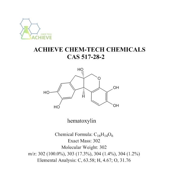


Shaanxi BLOOM Tech Co., Ltd. is one of the most experienced manufacturers and suppliers of hematoxylin powder cas 517-28-2 in China. Welcome to wholesale bulk high quality hematoxylin powder cas 517-28-2 for sale here from our factory. Good service and reasonable price are available.
Hematoxylin powder, a natural dye with CAS 517-28-2 and molecular formula C16H14O6, is a pigment extracted from the dried branches of Haematoxylon campechianum in South America using ether. It is one of the most commonly used dyes and a natural medium dye. Used for dyeing silk, fur, and leather, as well as printing cotton fabrics, different mordants can be used to obtain blue or black colors. In aqueous solutions, especially in alkaline solutions, it is easily oxidized by air to form reddish brown oxidized hematoxylin. Hematoxylin cannot be directly stained and must be exposed to a well ventilated area to turn it into oxidized hematoxylin (also known as hematoxylin) before it can be used, which is called "maturation".

|
Chemical Formula |
C16H14O6 |
|
Exact Mass |
302 |
|
Molecular Weight |
302 |
|
m/z |
302 (100.0%), 303 (17.3%), 304 (1.4%), 304 (1.2%) |
|
Elemental Analysis |
C, 63.58; H, 4.67; O, 31.76 |
The maturation process of hematoxylin takes a long time, and the longer it is prepared, the stronger its staining power. The dyed material must undergo the action of a metal salt as a mediator before it can have coloring power. So when preparing hematoxylin dye, a mordant must be used. Common mordants include aluminum sulfate, potassium alum, and iron alum. Hematoxylin is a light yellow to rust purple crystalline substance that is insoluble in cold water, ether, and glycerol, easily soluble in hot water and hot alcohol, and soluble in solutions of alkali, ammonia, and borax. It is an excellent material for staining cell nuclei and can differentiate different structures in cells into various colors.
|
|
|


The color of the tissue stained during differentiation varies depending on the treatment. After differentiation with an acidic solution (such as hydrochloric acid alcohol), it turns red, and after washing with water, it still returns to bluish blue. After differentiation with an alkaline solution (such as ammonia water), it turns blue, and after washing with water, it turns blue black. Mainly causing the chromatin in the nucleus and ribosomes in the cytoplasm to appear purple blue; Eosin is an acidic dye that primarily causes components in the cytoplasm and extracellular matrix to turn red. HE staining is the most fundamental and widely used technique in teaching and research of histology, embryology, and pathology.

Hematoxylin powder is a natural polyphenolic alkaline dye extracted from the heartwood of the legume plant Eucommia ulmoides. Its core staining active ingredient is hematoxylin red, which is formed by oxidation and maturation. With its strong affinity for acidic nucleic acids in the chromatin of the cell nucleus, stable staining color, and strong compatibility with various counterstaining agents, it has become the most widely used classic staining reagent in the field of histology and pathological diagnosis in the past 150 years. It is an irreplaceable core component in the conventional HE staining system.
Unlike artificially synthesized aniline dyes, the nucleus of hematoxylin stained cells presents a uniform and transparent blue purple color with delicate color layers. The subtle textures of nucleoli and chromatin can be clearly presented, and it can adapt to almost all types of biological samples from paraffin sections, frozen sections to cell smears, covering dozens of sub scenarios such as clinical pathological diagnosis, biological research, forensic identification, etc. It is a core basic reagent supporting the establishment and development of modern tissue pathology systems.

Application of nuclear core staining in routine HE staining for clinical pathology
The most core and widely used use of hematoxylin is as a nuclear exclusive staining agent for routine HE staining in clinical pathology, and it is the most commonly used staining reagent in all hospital pathology departments on a daily basis. In the standard staining process of conventional paraffin sections, tissue sections that have undergone dewaxing and hydration are immersed in mature hematoxylin staining solution that has been stained with alum for 3-5 minutes.
The chromatin in the cell nucleus specifically binds to the hematoxylin aluminum mordant complex and is stained uniformly deep blue purple. Subsequently, after eosin counterstaining, the cytoplasm and intercellular matrix present a bright pink color. The strong color contrast between blue purple and pink makes the morphological boundaries, nuclear cytoplasmic ratio, and tissue structure levels of the cells clear and distinguishable. Pathologists can easily identify the core morphological basis for benign and malignant judgments such as cell heterogeneity and nuclear division.


Clinical statistics from the pathology departments of top tier hospitals in China show that over 95% of routine pathological diagnoses rely on HE staining, and the quality of hematoxylin staining directly determines the final quality of HE sections. High quality hematoxylin staining can make nucleolar structures with a diameter of only 1 μ m clear and distinguishable, greatly reducing the risk of missed diagnosis of early-stage microcarcinoma. In the rapid staining scenario of intraoperative frozenpathology, the improved rapid hematoxylin powder staining solution can compress the staining time of cell nuclei to less than 1 minute. The contour of the stained frozen section cell nucleus is sharp, without the common problems of blurry cell nuclei and gray background in traditional rapid staining.
Fully meeting the requirement of rapid imaging of diagnostic results within 30 minutes of intraoperative pathology. In the pathological screening scenario of primary medical institutions, the stability advantage of hematoxylin staining is further highlighted. Qualified mature hematoxylin staining solution can be reused for several months at room temperature, and the staining effect will not show significant attenuation. It does not require expensive fully automatic staining equipment, and only basic staining instruments are needed to complete the preparation of qualified pathological sections.


It perfectly adapts to the limited resources of primary pathology laboratories and provides a low-cost and highly reliable staining solution for large-scale cancer early screening work. Another major advantage of hematoxylin staining is that the stained sections have extremely strong light resistance, and there will be no obvious fading of the cell nucleus even after being placed in a natural indoor environment for decades. The HE sections archived by the hospital pathology department from decades ago can still clearly observe the morphology of the cell nucleus, fully meeting the industry standard requirements for long-term archiving and retrospective of pathological data. This is a characteristic that the vast majority of artificially synthesized cell nucleus dyes cannot achieve.
Exclusive staining use for special tissue chemistry and target structures
In addition to conventional nuclear staining, hematoxylin has been modified with different mordant formulations to derive dozens of special staining systems, achieving targeted recognition of specific tissue components and pathological structures, and playing an irreplaceable role in pathological special diagnostic scenarios. In the context of Nissl staining in neural tissue, the modified toluidine blue hematoxylin combined staining system using alum hematoxylin can specifically stain Nissl in the cytoplasm of neurons into deep blue.


Clearly presenting the distribution and density changes of Nissl in the cytoplasm of neurons. This helps neuropathologists accurately identify the pathological changes of neurons in nerve injury and neurodegenerative diseases, and has been widely used in the pathological research and clinical diagnosis of Alzheimer's disease. In the context of striated muscle staining in muscle tissue, the phosphotungstic acid hematoxylin (PTAH) special staining system is a classic special staining method derived from hematoxylin. The combination of hematoxylin and phosphotungstic acid in this formula can specifically stain the transverse stripes of skeletal muscle and the intercalated disc structure of myocardium into a deep blue black color.
At the same time, the collagen fibers and cellulose components in the diseased tissue can be clearly distinguished and stained, providing highly recognizable morphological evidence in the pathological diagnosis of rhabdomyosarcoma and myocardial lesions. In the context of bile duct structure recognition in liver pathology, the improved Mayer hematoxylin low concentration long-term staining method can specifically outline the cytoplasm and luminal structure of bile duct epithelium without clearly staining the nucleus, helping pathologists accurately identify subtle lesions such as small bile duct hyperplasia and bile duct injury in liver biopsy tissue.


And providing key morphological support for the pathological diagnosis of autoimmune liver disease. In the scenario of staining decalcified sections of bone tissue, hematoxylin can tolerate the weakly acidic environment of residual decalcifying solution well. As long as it undergoes appropriate cyanation treatment, it can still clearly stain the nuclei of bone cells and the cell boundaries of bone trabeculae, without the problem of complete loss of staining in acidic environments like many artificially synthesized dyes. It is the most reliable nuclear staining reagent in the pathological diagnosis of bone tumors.
In the scene of acid fast staining and re staining of granulomatous lesions, after finding red tubercle bacillus through acid fast staining, dilute hematoxylin powder is used for light re staining, which can make the nucleus in the background appear light blue purple, and the identification of tubercle bacillus in the light blue background is greatly improved, effectively reducing the visual fatigue of pathologists looking for tubercle bacillus under the microscope, and improving the positive detection rate of tuberculosis pathology.


Application of immunohistochemistry and molecular pathology in nuclear counterstaining
In the context of molecular pathology techniques such as immunohistochemistry staining and in situ hybridization for modern precision pathological diagnosis, hematoxylin is the most widely used nuclear counterstaining reagent and an indispensable background staining component in all immunohistochemical sections. The core goal of immunohistochemical staining is to display positive signals of specific target proteins. The most commonly used DAB staining system will make the positive signal appear brownish yellow.
At this time, light stained hematoxylin is used to counterstain the cell nucleus, making the negative cell nucleus that does not express the target protein appear uniformly light blue. The brownish yellow positive signal has a clear boundary under the light blue cell nucleus background. Pathologists can accurately determine whether the positive signal is located in the nucleus, cytoplasm, or cell membrane, and accurately complete the interpretation and semi quantitative scoring of immunohistochemical results. In the interpretation of immunohistochemical indicators such as PD-L1 and Ki-67 related to tumor targeted therapy, the quality of hematoxylin counterstaining directly determines the accuracy of positive cell counting.


The uniform and light blue nuclear background allows pathologists to easily distinguish tumor cells from stromal cells, accurately calculate the proportion of positive tumor cells, and provide precise pathological basis for targeted and immunotherapy drugs for tumor patients. In the molecular pathology detection scenario of fluorescence in situ hybridization (FISH), hematoxylin serves as a bright field pre staining reagent, which can clearly outline the cell contour of the tissue before fluorescence observation, helping experimenters quickly locate the target tumor cell area, greatly improving the observation efficiency of FISH probe hybridization signals.
Avoiding ineffective search for target cells in a large number of interstitial cells, and significantly improving the efficiency and accuracy of FISH detection. In special immunoelectron microscopy sample preparation scenarios, the uranyl acetate hematoxylin combined staining method can enhance the electron density of cell membranes and nuclear membranes in ultra-thin sections, making the cellular ultrastructure layer clearer under electron microscopy, and helping pathological researchers accurately observe the morphology of organelles and subcellular localization of viral particles inside cells.


It has been widely used in ultra-thin pathological research in virology and neuroscience. In the liquid based thin-layer cell diagnosis scenario of clinical cytology, hematoxylin powder staining can clearly present the chromatin texture of cervical exfoliated cells, and pathologists can accurately identify the subtle morphological characteristics of irregular nuclear membrane and uneven distribution of chromatin in the nucleus, greatly improving the positive detection rate of cervical cancer liquid based cytology screening and reducing the risk of false negative and missed diagnosis.
FAQ
Is hematoxylin pink or purple?
Hematoxylin and Eosin, commonly reffered to as H&E, is considered a routine stain in identifying the morphological structures within a tissue section. The nuclei in this stain are stained a blue-purple hue from the hematoxlyin. Eosin is used as a counter stain to mark cytoplasm and muscle fibers in varying pink hues.
What is a substitute for hematoxylin?
The iron alum-celestine blue solution is stable for a few months. A staining time of 5-10 minutes is usually satisfactory for formalin fixed tissues. This method has been recommended as a substitute for Hematoxylin and Eosin.
What is the common name for hematoxylin?
As a dye, haematoxylin has been called palo de Campeche, logwood extract, bluewood and blackwood. In histology, haematoxylin staining is commonly followed by counterstaining with eosin. When paired, this staining procedure is known as H&E staining and is one of the most commonly used combinations in histology.
Is hematoxylin positive or negative?
Hematoxylin in complex with aluminum is cationic and acts as a basic dye. Because it is positively charged, it can bind to negatively charged, basophilic cell components, such as nucleic acids in the nucleus. These get stained blue or purple as a result.
Hot Tags: hematoxylin powder cas 517-28-2, suppliers, manufacturers, factory, wholesale, buy, price, bulk, for sale, Indicator Reagent, Phenol red powder, 2-Iodoxybenzoic Acid CAS 61717-82-6, Dithizone Reagent, p-Dimethylaminobenzaldehyde, 4 5 Chloro 2 pyridylazo 1 3 phenylenediamine